
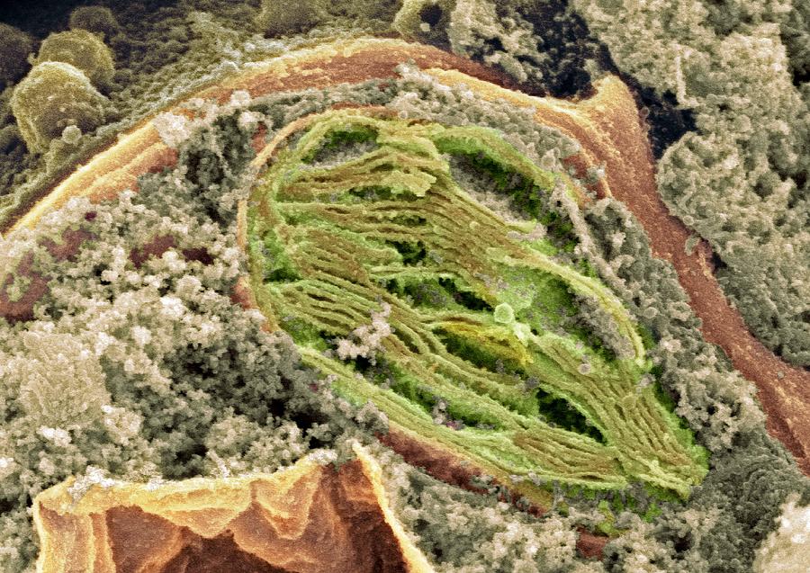
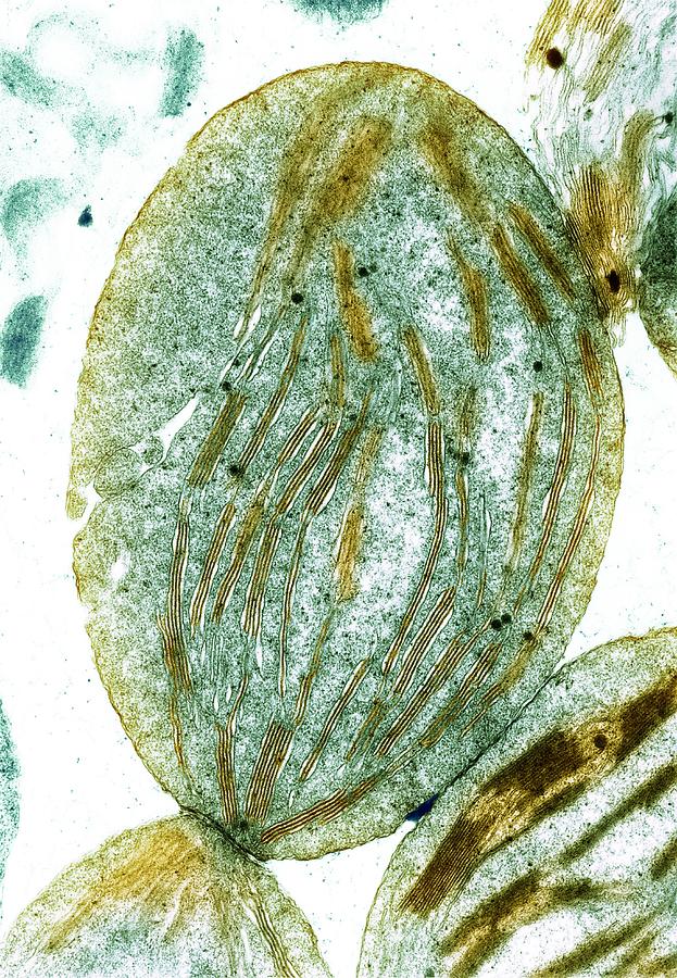
Tem Of Chloroplasts Photograph by Dr Jeremy Burgess
Serial-Block-Face Scanning Electron Microscopy (SBF-SEM) associated with biomolecular analysis show that chloroplast differentiation proceeds by distinct 'structure establishment' and 'chloroplast proliferation' phases, each with differential protein and lipid regulation.

ISOLATION OF CHLOROPLAST Bioscience Notes
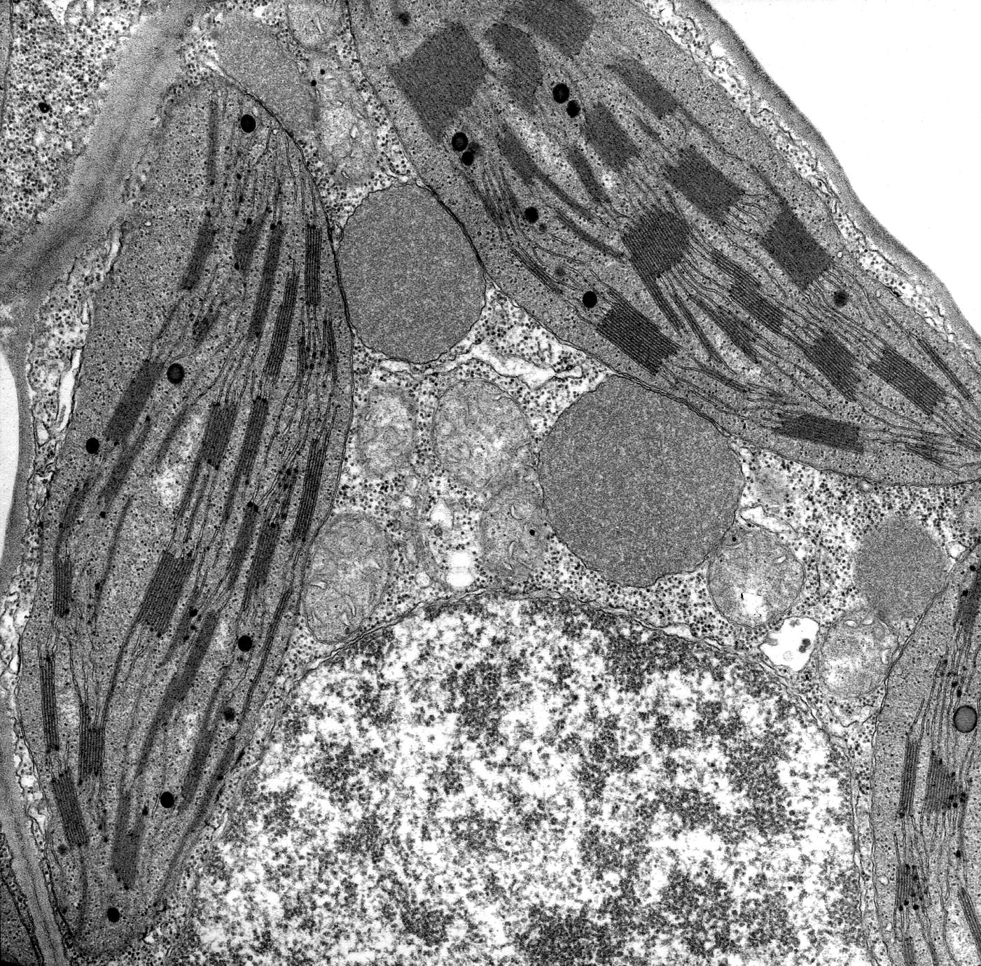
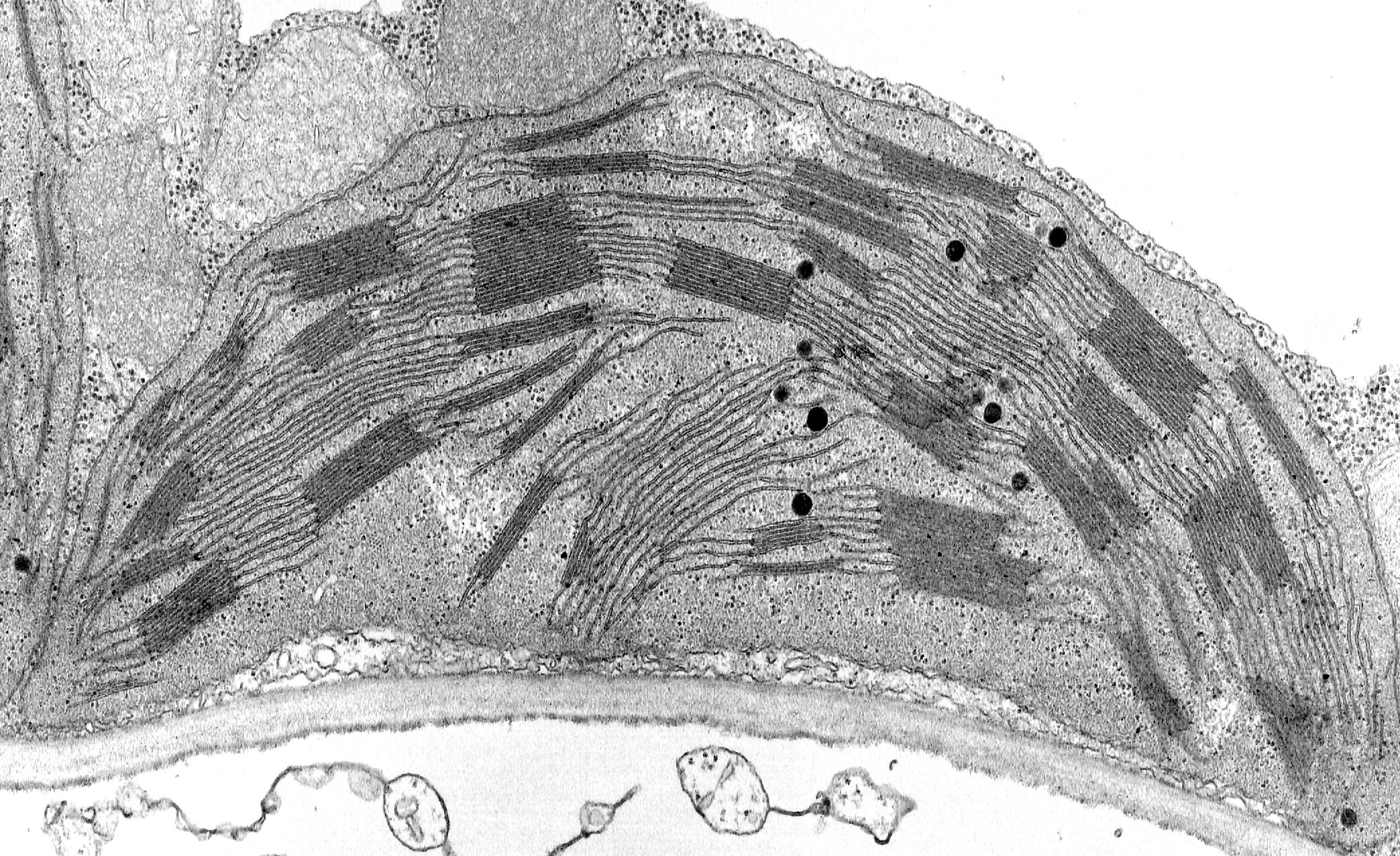
Figure 23 (a) Electron micrograph of a chloroplast. A: double outer membrane or envelope (the outer and inner membranes are labelled separately in (b)); B, stroma; C, stroma lamellae; D, granum, composed of a stack of thylakoids. (b) Schematic diagram of chloroplast structure. Show description Figure 23
Chloroplast, Sem Photograph by Dr David Furness, Keele University Fine Art America
Chloroplast nucleoids are thought to be the functional unit for various processes, including DNA replication, repair, recombination, inheritance, and transcription, and are often compared with.
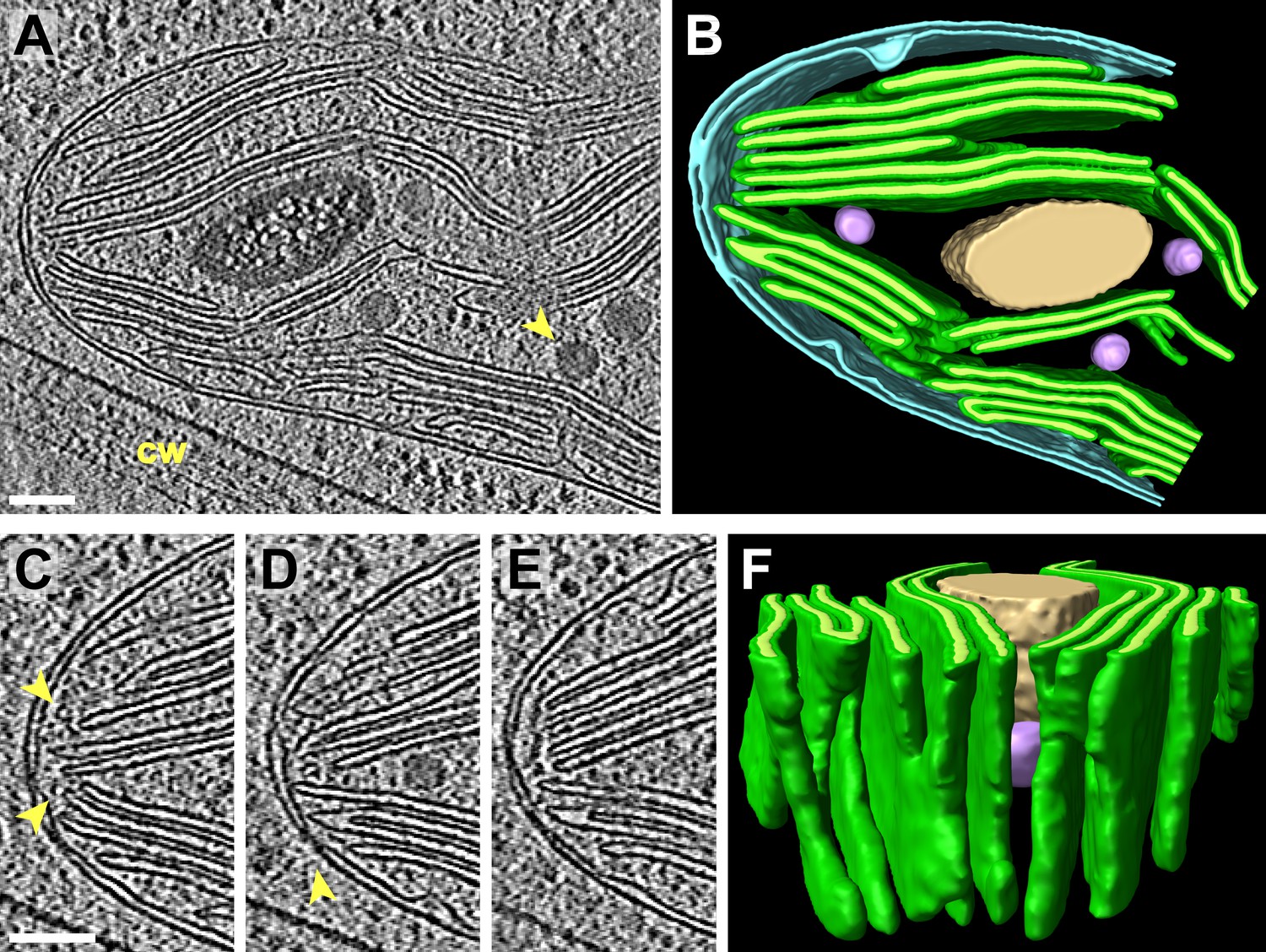
Native architecture of the Chlamydomonas chloroplast revealed by in situ cryoelectron
Chloroplasts in C4 plants exhibit structural dimorphism because thylakoid architectures vary depending on energy requirements. Advances in electron microscopy imaging capacity and sample preparation technologies allowed characterization of thylakoid structures and their macromolecular arrangements with unprecedented precision mostly in C3 plants.
Nucleus, glyoxisomes, chloroplasts, and mitochondria magnification at 13,900x UWDC UW
Cellular Component. chloroplast. Electron micrograph of a thin section through a portion of the chloroplast of the unicellular green alga Chlamydomonas. Free ribosomes are seen as dark particles within the chloroplast stroma. The ima. CIL:41049. NCBI Organism Classification.

TEM of chloroplasts from a pea plant Stock Image B110/0043 Science Photo Library
The thylakoid architecture of such morphologically variable chloroplasts is confirmed by transmission electron microscopy (TEM). The method of monitoring structural variation by light microscopy in combination with electron microscopy is described. Keywords: Light microscopy, TEM, Chloroplast, Thylakoid membrane, Arabidopsis thaliana Go to:
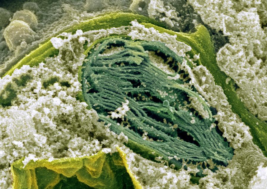
Chloroplast, Sem Photograph by Dr David Furness, Keele University Fine Art America
Chloroplasts are found only in plants and photosynthetic algae. (Humans and other animals do not have chloroplasts.). Electron micrograph of a mitochondrion, showing matrix, cristae, outer membrane, and inner membrane. _Image credits: upper image, "Eukaryotic cells: Figure 7," by OpenStax College, Biology . Modification of work by Matthew.
Chloroplast, Sem Photograph by Dr David Furness, Keele University Fine Art America
The chloroplast organelle in mesophyll cells of higher plants represents a sunlight-driven metabolic factory that eventually fuels life on our planet. Knowledge of the ultrastructure and the dynamics of this unique organelle is essential to understanding its function in an ever-changing and challenging environment.

Chloroplasts, light micrograph Stock Image C011/9275 Science Photo Library
Chloroplasts The sketch of the chloroplast above was made from an electron micrograph of a chloroplast from a higher order plant (Levy).. both photosystems are used in an electron transport process that yields energy in the form of ATP and reduced coenzymes to the stroma of the chloroplast to be used in the synthesis of carbohydrates.
A. Electron microscopy picture of a higher plant chloroplast. B. Light... Download Scientific
Chloroplasts are the organelles specialized in carrying out the photosynthetic process, which uses light energy to synthesize organic compounds; for this reason, they are common to all photoautotrophic eukaryotes.
Chloroplast UWDC UWMadison Libraries
Chloroplasts, the organelles responsible for photosynthesis, are in many respects similar to mitochondria.. (Electron micrograph . Despite this greater complexity, the membranes of chloroplasts have clear functional similarities with those of mitochondria—as expected, given the role of both organelles in the chemiosmotic generation of ATP.
Transmission electron microscopy picture of the chloroplast envelopes.... Download Scientific
An electron micrograph showing the structures within a chloroplast Exam Tip Make sure you can identify the structures of a chloroplast on a diagram AND that you can explain the function of each of these structures. You've read 1 of your 10 free revision notes Get unlimited access to absolutely everything: Downloadable PDFs Unlimited Revision Notes
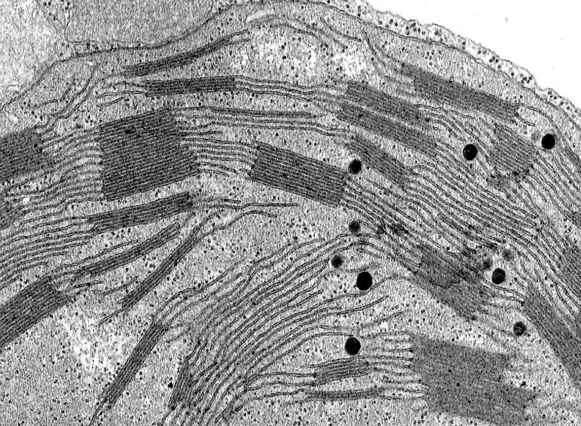
Detail of a chloroplast with well developed grana UWDC UWMadison Libraries
chloroplast, structure within the cells of plants and green algae that is the site of photosynthesis, the process by which light energy is converted to chemical energy, resulting in the production of oxygen and energy-rich organic compounds.
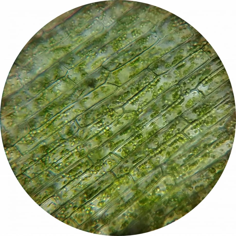
microscopeview of plant chloroplast free image Peakpx
As reviewed by Staehelin in 2003, the study of chloroplast structure has advanced with enhancements in light and electron microscopy from two-dimensional transmission electron microscopy (TEM) imaging to three-dimensional (3D) electron tomography (ET) and with improvements in sample preservation from chemically preservation to freeze-fracture to.

TEM of chloroplast from Coleus blumei Stock Image B110/0067 Science Photo Library
Download scientific diagram | Electron micrograph of a chloroplast in a field-collected Elysia clarki. The thylakoids (TH) are arranged in bands of two to six lamellae all running in the same.

Electron micrograph of a chloroplast in a fieldcollected Elysia... Download Scientific Diagram
Electron micrograph of a chloroplast The inner membrane system of the chloroplast is called the thylakoid membranes and the matrix surrounding the thylakoids is called the stroma. Stacks of thylakoids are termed the grana, while the membranes connecting the grana are called the stroma thylakoids.
